
London Vet Show 2022
No foot, no horse - how to take and interpret foot radiographs
No foot, no horse - how to take and interpret foot radiographs
Couldn't load pickup availability
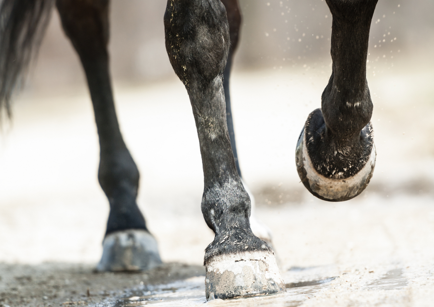
Diseases of the foot are one of the most common sources of lameness in horses. Due to its relative low cost and wide availability, radiographic examination of this area is one of the first instance to diagnose potential abnormalities of this area. However, a radiographic diagnosis can only be made on good quality radiographs. Correct and careful positioning of the horse as well as knowledge of the radiographic equipment are crucial for optimal images. For recognising lesions, familiarity with the normal anatomy is as important as knowledge of radiographic appearance of abnormalities such as disease of the distal interphalangeal joint, navicular bone and other structure of the equine foot. This talk aims to help practitioners to take good quality radiographs of the equine foot and recognise common and not so common abnormalities of this area.
- •How to take good quality radiographs of the equine foot
- •Assessing and improving the diagnostic quality of radiographs
- •Recognise common and not so common abnormalities of equine foot
Presented at London Vet Show 2022
Thursday 17th November 2022 10:20
RVC Equine Theatre 1
Please note this session is not RACE-approved but you can still earn a CPD certificate
Share